Sarcomere simulation: observe the sliding of actin and myosin filaments by changing the contraction rate; measure that the A band remains constant while I and H bands shorten; order the contraction steps and examine the muscle twitch curve
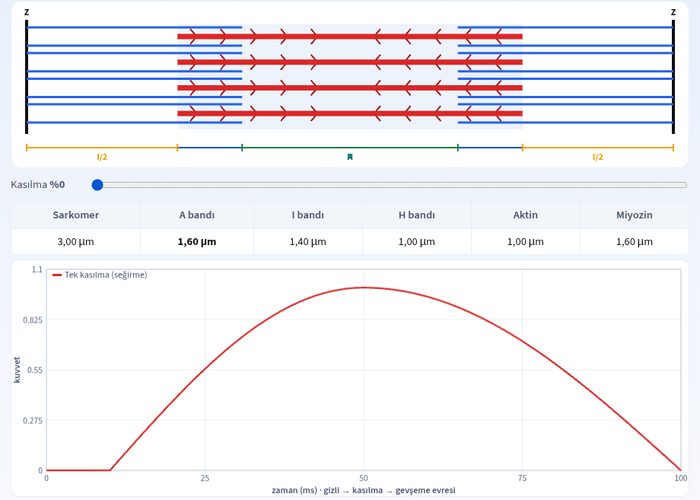
Interactively explore the structure of the sarcomere and the sliding filament model with the skeletal muscle contraction and relaxation simulation. In this experiment, by changing the contraction rate, you will watch the actin and myosin filaments slide relative to each other and observe the changes in the A band, I band, and H band. It is a great resource to reinforce muscle physiology topics in biology class.
What can you do in this simulation?
You can examine filament movements by changing the contraction rate on the sarcomere simulation.
You can measure the dimensional changes of the A band, I band, and H band under full contraction and full relaxation conditions.
You can sequence the contraction steps from the release of acetylcholine by the motor neuron to the shortening of the sarcomere.
You can test the stages of the sliding filament model with interactive cards and questions.
Basic concepts and formulas
The structural unit of skeletal muscle is the sarcomere. During contraction, actin filaments slide over myosin filaments and approach each other. In this process, the length of the sarcomere shortens, the I band and H band narrow or disappear, but the length of the A band, which represents the length of the myosin filaments, does not change. Ca²⁺ ions and the ATP molecule are mandatory for contraction.
How to use?
Go to the simulation panel and examine the structure of the sarcomere with the guidance.
Observe the movement of filaments and changes in the bands by adjusting the contraction rate.
Drag and drop the cards to arrange the contraction steps in the correct order.
Try your predictions by answering the questions and check your score.
Frequently asked questions
Why does the length of the A band not change during contraction?
The A band represents the length of the myosin filaments. As actin slides over myosin, there is no change in the length of the myosins, which is why the A band remains constant.
Why do the I and H bands shorten during contraction?
Because actin filaments are pulled toward the center of the myosin, the two Z lines move closer to each other, narrowing the I band formed by actin and the H band, which is the gap in the middle of the myosins.
Why is ATP required for contraction?
Energy is required for myosin heads to bind to actin, pull the filaments (power stroke), and subsequently detach. If ATP is depleted, myosin heads cannot detach from actin, and the muscle remains contracted.
 Interactive experiment